
News
Sometimes the most valuable scientific discoveries come not from proving that something works, but from understanding where its limitations lie…
In a recent publication supported, in part, by the DCHSB Computational Core entitled, "Challenges of Conventional Iterative All-Atom and Coarse-Grained Multiscale Molecular Dynamics," where Dr. Hung N. Do, Joe McKenzie, and Dr. S. Gnana Gnanakaran, examined a question that many computational biologists have considered but that has received surprisingly little systematic evaluation: Can we (collectively) improve biomolecular sampling by repeatedly cycling between all-atom and coarse-grained representations? To address this, they developed an iterative multiscale molecular dynamics (iMMD) workflow available on github, and tested it across diverse systems ranging from soluble proteins to complex membrane proteins. One example shown below was from figure 5 of the paper.
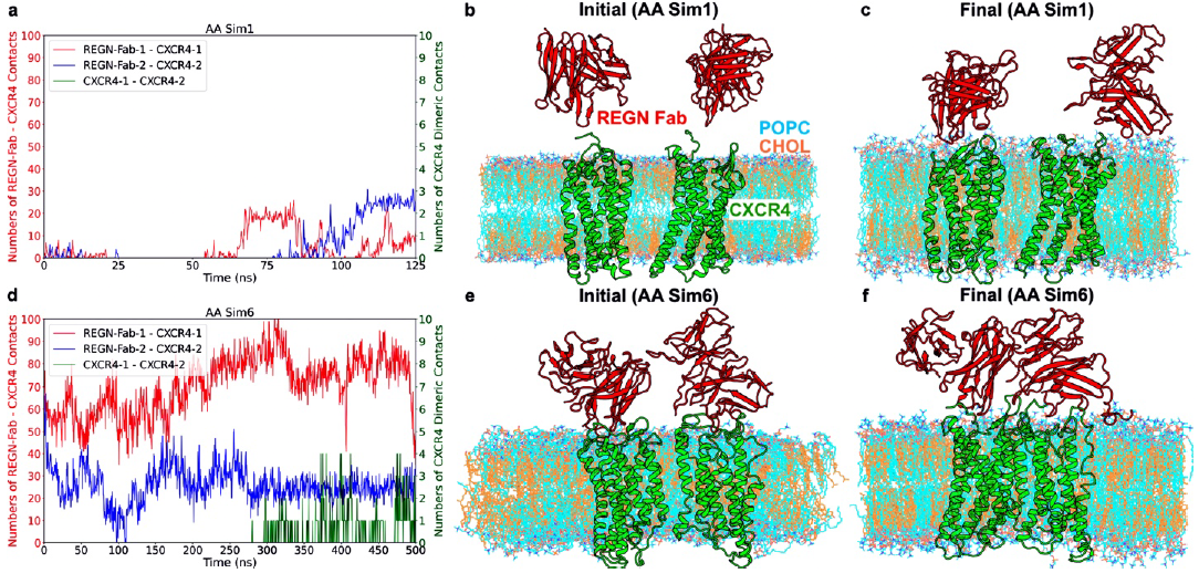
Figure 5. Binding of the REGN7663 Fabs to dimerizing CXCR4 receptors in the heterogenous CHOL/POPC lipid bilayer observed during iMMD simulations. (a) Time courses of the numbers of contacts between the two CXCR4 receptors and between the REGN Fabs and the CXCR4s calculated from the first AA MD simulation iteration. The left y-axis is colored red and represents the number of contacts between the REGN Fabs and CXCR4s, while the y-axis is colored green and represents the number of contacts between the two CXCR4s.(b-c) The initial (b) and final (c) conformations of the REGN Fabs and CXCR4s in the CHOL/POPC lipid bilayer obtained from the first AA MD simulation iteration. (d) Time courses of the number of contacts between the two CXCR4 receptors and between the REGN Fabs and the CXCR4s calculated from the sixth AA MD simulation iteration. (e-f) The initial (e) and final (f) conformations of the REGN Fabs and CXCR4s in the CHOL/POPC lipid bilayer obtained from the sixth AA MD simulation iteration.
Their finding reveal both opportunities and caveats and remind us that understanding what does not work and why can be just as valuable as discovering what does. We hope these findings help guide the development of more robust multiscale simulation approaches for studying biomolecular structure, dynamics, and function at increasingly realistic scales.
Use this link to download the full pdf.
Our collaborative teams in DCHSB uncovered a mechanism that explains how HIV-1 detects its host cell and initiates the structural rearrangements necessary for host-cell entry.
Capturing how HIV-1 transitions from receptor engagement to membrane fusion has been one of the major challenges in structural virology. By combining cryo-EM, molecular simulations, virology, and structural biology, our collaborative team uncovered a mechanism that explains how HIV-1 detects its host cell and initiates the structural rearrangements necessary for host-cell entry.
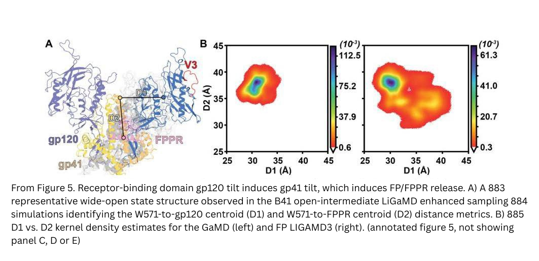
The recent publication from studies lead by Drs. Rory Henderson and S. Gnana Gnanakaran’s labs entitled, “Sequential HIV-1 Fusion Protein Gates Control the Initiation of Host Cell Entry” is now available as a preprint on BioRxiv. Lead authors Drs. Hung Do (NIH) and Muralikrishna Lella along with co-authors, Devansh Fulmali, Niva Rajpara, Ilona Unarta, Alexis Johnson, Katarzyna Janowska, Helena Laukaitis, Carrie Saunders, R.J. Edwards, Priyamvada Acharya, Amit Sharma, S. Gnanakaran, and Rory Henderson, identified two sequential conformational “gates” within the HIV-1 Envelope (Env) fusion machinery that regulate fusion peptide release and subsequent gp120 shedding, key steps required for successful infection.
This project highlights the value of bringing together complementary expertise across institutions and disciplines, including structural biology, cryo-electron microscopy, computational biophysics, molecular simulation, virology, and immunology. The collaboration between teams at Duke Human Vaccine Institute, Los Alamos National Laboratory, and North Carolina State University enabled us to connect molecular structures, dynamics, and biological function into a unified model of HIV-1 entry.
Use this link to download the full pdf.

Congratulations Dr. Ahn!
Congratulations on the 2024 Small Pilot Awards!
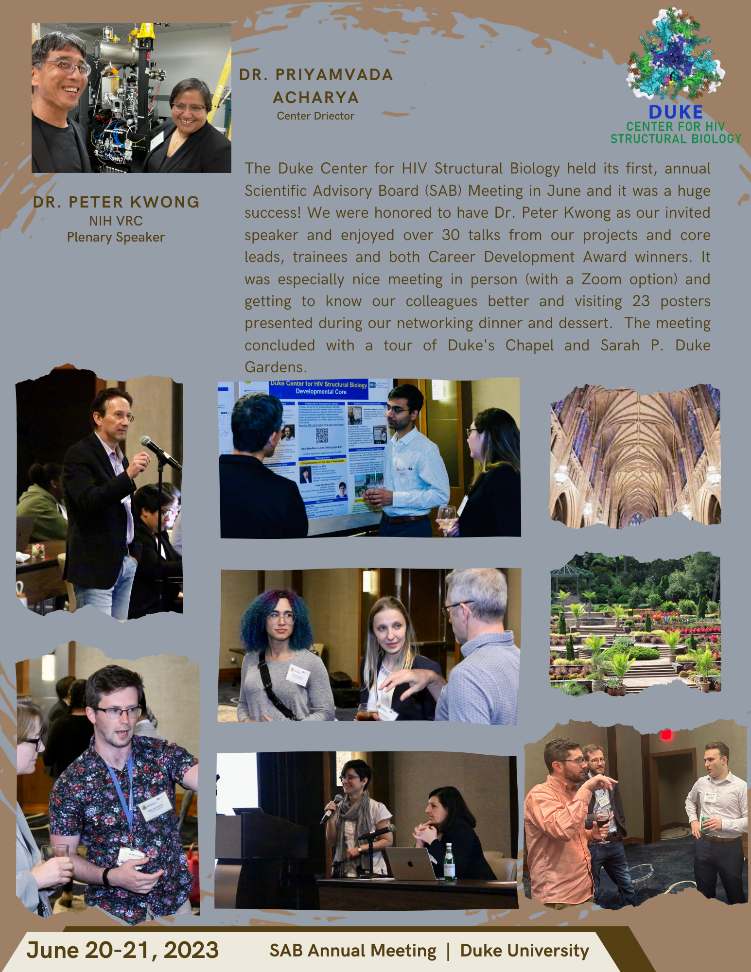
2023 SAB Annual Meeting
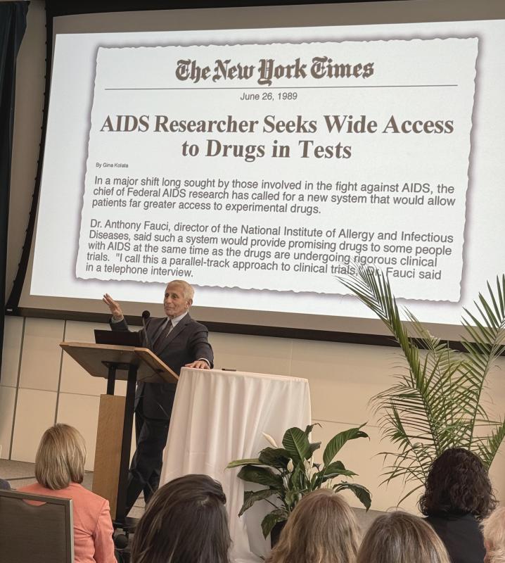
DHVI Co-Hosts Norman L. Letvin, MD, Memorial Lecture Featuring Anthony Fauci, MD, at Invitation of Barton Haynes
Anthony S. Fauci, MD, was chief of the Laboratory of Immunoregulation at the National Institute of Allergy and Infectious Disease (NIAID) when he read about a strange form of pneumonia that had killed more than two dozen young, otherwise healthy gay men in Los Angeles in the span of a month. Continued
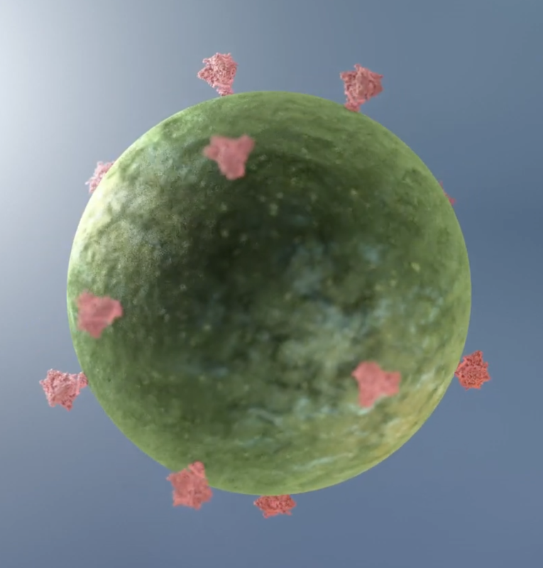
A Trial HIV Vaccine Triggered Elusive and Essential Antibodies in Humans
An HIV vaccine candidate developed at the Duke Human Vaccine Institute triggered low levels of an elusive type of broadly neutralizing HIV antibodies among a small group of people enrolled in a 2019 clinical trial.
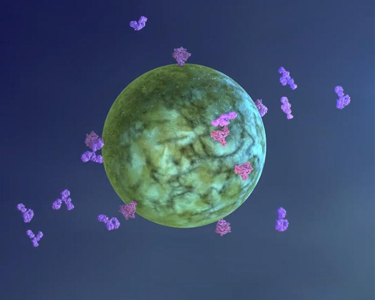
GPS-like System Shows Promise as HIV Vaccine Strategy to Elicit Critical Antibodies
DURHAM, N.C. – A team led by the Duke Human Vaccine Institute (DHVI) has developed a vaccine approach that works like a GPS, guiding the immune system through the specific steps to make broadly neutralizing antibodies against HIV.

Duke Human Vaccine Institute Wins Contract to Produce Pan-Coronavirus Vaccine
The Duke Human Vaccine Institute has received a federal contract to manufacture a pan-coronavirus vaccine candidate that can be tested in a phase 1 clinical trial.
Awarded by the National Institute of Allergy and Infectious Diseases (NIAID), part of the National Institutes of Health (NIH), the base period of the contract provides $11.2 million to support the program; additional provisions in the contract could increase the total funding up to $21.5 million if all option periods are exercised.

Duke Awarded Federal Grant to Build Structural Models of HIV
Researchers at the Duke Human Vaccine Institute (DHVI) received a federal grant totaling more than $27 million over five years to focus on building structural models of HIV that will help guide the development of therapies and vaccines.
Announcements
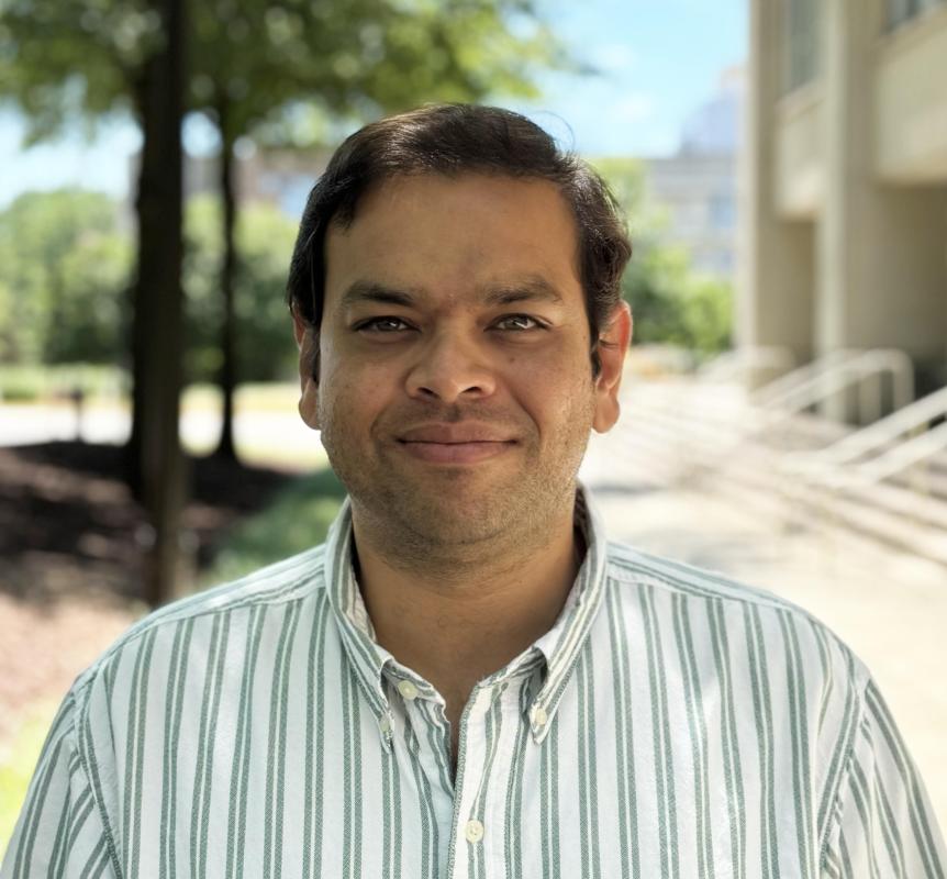
DHVI Faculty Appointments, Promotion, and Tenure - Kshitij Wagh, PhD
The Duke Human Vaccine Institute is pleased to announce that Kshitij Wagh, PhD, has been recruited to a faculty position in the Duke Human Vaccine Institute (DHVI), effective May 15, 2024.